Locations

Fax: (303) 762-9292

Computer Enhanced Navigation: Image Guided Spine Surgery

Image Guided Surgery or IGS technology and theory is complex. However, the key fact to remember is this technology has a profound impact on ensuring better and safer spinal surgery outcomes.
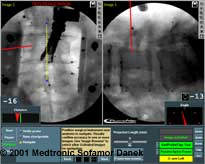
Image guidance technology creates three-dimensional (3D) models of the spine and virtual images of the surgeon’s instruments during the operation. The images can be manipulated and merged to reveal greater detail when planning or performing surgery. Surgeons can navigate the spine’s complex anatomy, visualize disorders, and perform tasks more accurately such as implanting spinal instrumentation.

What’s Involved - How it Works
To bring this technology to life involves a high-performance computer, sophisticated software, monitor, camera(s) to recognize light emitting diodes (LEDs), and instruments fitted with LEDs.
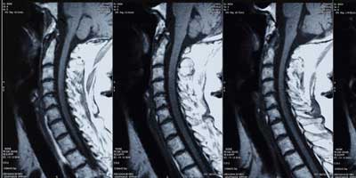
Days before surgery, CT scans and/or MRI images of the patient’s anatomy are loaded into the computer and 3D images are created. The images can be rotated, enlarged (zoom feature), or manipulated in different ways allowing the surgeon to pre-plan the patient’s surgery. As a pre-operative tool, the surgeon can predetermine the type and size of instrumentation needed and even plot the trajectory of pedicle screws.
During surgery, the surgeon sees the operation on the monitor because the instruments fitted with LEDs communicate with the computer in real time. In other words, the surgeon sees the position of the instruments as it relates to the anatomy, even if the anatomical area is hidden from the surgeon’s direct view.
Advantages
Our surgeons find image guided surgery is beneficial because:
- It enhances their ability to navigate complex spinal anatomy in real time.
- It provides accurate measurements, such as screw diameter and length.
- It allows more precise implantation of spinal instrumentation.
- It can pinpoint exactly where the tip of a surgeon’s instrument is in relation to a patient’s anatomy, providing invaluable information in case unexpected or unusual anatomy is encountered.
- It reduces operating time.
- It minimizes (and in some cases, eliminates) radiation exposure previously experienced with traditional x-rays.
Conclusion
While image guidance technology (IGS) is remarkably complex, it has a very simple benefit: enhanced patient safety. Patients can rest assured that the very latest technology is being utilized to ensure the best possible surgical outcomes.


