Locations

Fax: (303) 762-9292

Understanding Spinal Anatomy: Spinal Cord and Nerve Roots
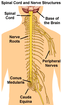
The spinal cord is a slender cylindrical structure about the diameter of the little finger. The spinal cord begins immediately below the brain stem and extends to the first lumbar vertebra. Thereafter, the cord blends with the conus medullaris which becomes the cauda equina, a group of nerves resembling the tail of a horse.

The spinal nerve roots are responsible for stimulating movement and feeling. The nerve roots exit the spinal canal through the intervertebral foramen, small hollows between each vertebra.
The brain and the spinal cord make up the Central Nervous System (CNS). The nerve roots that exit the spinal cord/spinal canal branch out into the body to form the Peripheral Nervous System (PNS).
| Type of Neural Structure | Role/Function |
|---|---|
| Brain Stem | Connects the spinal cord to other parts of the brain. |
| Spinal Cord | Carries nerve impulses between the brain and spinal nerves. |
| Cervical Nerves (8 pairs) | These nerves supply the head, neck, shoulders, arms, and hands. |
| Thoracic Nerves (12 pairs) | Connects portions of the upper abdomen and muscles in the back and chest areas. |
| Lumbar Nerves (5 pairs) | Feeds the lower back and legs. |
| Sacral Nerves (5 pairs) | Supplies the buttocks, legs, feet, anal and genital areas of the body. |
| Dermatomes | Areas on the skin surface supplied by nerve fibers from one spinal root. |

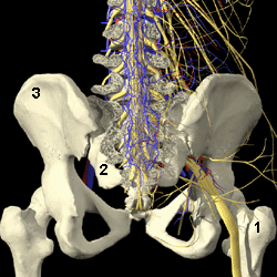
- Sciatic Nerve (yellow)
- Part of the Sacrum
- Hip Bone
Between the front and back portions of the vertebra (i.e. midregion) is the spinal canal that houses the spinal cord and the intravertebral foramen. The foramen are small hollows formed between each vertebra. These hollows provide space for the nerve roots to exit the spinal canal and to further branch out to form the peripheral nervous system.


